السلام عليكم ورحمة الله وبركاته
وجدت في هذا الموقع معلومات جد مهمة لكيفية فهم وقراءة مخطط الدم الكامل CBC اوComplete Blood Count
سواء كنت تعاني من فقر دم او التهاب معين تستطيع عن طريقة مخطط الدم الشامل معرفة قيمة خضاب الدم وهل تعاني من فقر دم Anemia اوارتفاع قيمة الكريات البيض Leucocytes او نقص في الصفيحات
اعتقد أن هذه المعلومات جدا ضرورية لكل انسان و ليس فقط الاطباء
A complete blood count (CBC):
is the most widely requested and single most important lab test on blood. Many CBCs are done as routine screens – tests that provide general information about the patient’s status. Most CBCs include the following cellular measurements:
•
WBC count
•
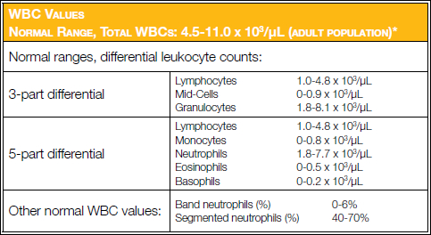
WBC differential, 3-part: lymphocytes, mid-cells, granulocytes
OR
5-part: lymphocytes, monocytes, neutrophils, eosinophils, basophils
• RBC count
• Hemoglobin (Hb)
• Hematocrit (Hct)
• RBC indices (MCV, MCH, MCHC)
• Platelet count
Normal ranges (also called reference ranges) depend on geographic location, the patient’s sex, and age; ranges are used as guidelines and vary based on specific populations.
White Blood Cell (Leukocyte) Count
This is a count of the number of WBCs present in a known volume of blood. Automated systems often count as many as 20,000 WBCs to ensure accuracy and precision. The WBC count is further identified by the major WBC sub-populations (WBC differential). WBC counts and the WBC differential are generally measured by optical technologies or a combination of optical, impedance, radio frequency, or multi-color fluorescence in modern hematology analyzers.
Less sophisticated hematology analyzers provide WBC sub-populations as 3-part WBC differentials reporting a percentage and absolute value for Iymphocyte, mid-cells, and granulocyte populations. Advanced hematology analyzers minimally provide a 5-part WBC differential reporting percentages and absolute values for lymphocytes, monocytes, neutrophils, eosinophils, and basophils. The modern hematology analyzer also indicates (flags) suspected abnormalities, from which the technologist may perform a microscopic smear review
or an actual manual differential count to ensure accuracy of the reported results.

WBC Reference Range (note:Reference ranges should be established by each laboratory.)
Red Blood Cell (Erythrocyte) Count. This is a count of the number of RBCs present in a known volume of blood. Automated systems often count as many as 20,000-50,000 RBCs to ensure precision. The RBC count is also used to calculate the RBC indices.

RBC count reference range
Hematocrit (Hct). This is the volume of RBCs in a whole blood sample expressed as a percentage (%).

Hematocrit (Hct) reference range
Hemoglobin (Hb). Hemoglobin values may be obtained in several ways. The most common method adds potassium cyanide (or similar compounds) to convert Hb to cyanmethemoglobin, which is measured with a spectrophotometer. The results, recorded as grams per deciliter (g/dL), indicate the oxygen-carrying capacity of the RBCs.

Hemoglobin reference range
RBC lndices
The RBC indices –mean cell volume (
MCV), mean cell hemoglobin (
MCH), and mean cell hemoglobin concentration (
MCHC) – indicate the volume and character of hemoglobin and, therefore, aid in the differential diagnosis of the type of anemia present.
Mean Cell Volume (MCV):
The MCV is the average volume of RBCs; it is calculated from the hematocrit (volume of packed red cells) and the erythrocyte count. This is the most important RBC index in the differential diagnosis of anemias. The value can be derived from the following equation:
MCV = Hct (%) x 10/RBC (x 10^6/μL)

MCV Reference range
Today’s modern hematology analyzers actually do cell sizing, therefore, the MCV is actually a measured parameter and Hct is calculated from the MCV.
Mean Cell Hemoglobin (MCH):
This is a calculation of the ratio of hemoglobin to the erythrocyte count. The formula for the calculation is:
MCH = Hb (g/dL)/RBC (x 10^6/μL) x 10

MCH Reference values
Mean Cell Hemoglobin Concentration (MCHC):
This calculation of the ratio of hemoglobin to hematocrit indicates the concentration of hemoglobin in the average red cell. The calculation is:
MCHC = [Hb (g/dL)/Hct (%)] x 100

MCHC Reference range
Reticulocyte Count:
For a count of these young RBCs, a few drops of blood are smeared on a slide, stained with methylene blue, and counterstained with Wright’s stain before being counted under a microscope. One thousand RBCs are counted and the number of cells having a blue-stained reticulum are expressed as a percentage. Now, most automated systems provide reticulocyte counts using various stains and dyes.

Reticulocyte Count
Platelet Count:
Because their normal numbers are so high, platelets were often estimated. Now, automated systems provide platelet counts from a variety of methods (i.e., impedance, optical, and immuno-platelet).

Platelets Count Reference Range


 رد مع اقتباس